
OSTEC ARTTOOL LIMITED COMPANY
Russia
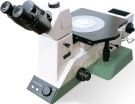
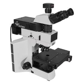
OMOS M series analytical metallographic systems make perfect solutions for the microstructure of materials automatic analysis. When you image and analyze samples, you are often faced with complex and interrupted processes that can make tasks take much longer than you want them to. OMOS M series microscopes have vast experience in bringing together optical precision, automation, analytical power, and data management via the workfloworiented system. The new M series analytical metallographic system product range takes this one step further, offering peerless flexibility and ease of use that can make any task a controlled workflow. Ostec is dedicated to ensuring that the best solutions are available for your work, from microscopes and digital cameras to software and data storage. OMOS M series microscopes bring all of our experience to you, giving you control over every aspect of your hardware, workflow
ADVANCED MAGNETIC TECHNOLOGIES AND CONSULTING (AMT&C)
Russia
The magnetic system of this field source is also built on the principle of nested cylindrical Halbach-structures. An important feature is the presence of two motors, controlled independently, so it is possible to manage the change not only the magnitude but also the direction of the magnetic field. Because the device was designed for operation together with an optical microscope, the vertical size of the system near the working region is reduced to 90 mm. Stepper motors, that turn the two magnetic subsystems, are located on the sides of the casing

CENTER OF INFORMATION TECHNOLOGIES NELIAN LLC
Russia
We advise you to order not the whole complete set of microscope + software Dianel®Micro + Digital camera + Set of consumables. We advise you to order only Dianel®Micro Software for Hemoscaning Diagnosis of health by a drop of live blood and Microscope Laboratory Researches (English version) + 0.01mm Reticule Calibration Slide Or Modernization Complete set of Dianelmicro software for hemoscaning + CCD video camera 3,2 Mpix matrix by Sony + 0.01mm Reticule Calibration Slide For you is much better to buy microscope in your country with all necessary certification than to import microscope from Russia. We will be very appreciate to help you to choose the optimal model of microscope for your needs. Our camera is compatible with almost all microscopes. But you can use your digital camera you already have or planning to purchase.

CENTER OF INFORMATION TECHNOLOGIES NELIAN LLC
Russia
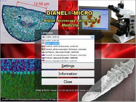
Diagnostic complex Dianel®Micro specal software “Dianel® micro” that compatible with most of all the digital microscope cameras. DianelMicro software is intended to visualize an image with a high resolution on the screen, automation of qualitative sample examination of any micro objects and bio materials, storage, measurement, analysis and systematization of photo images and video. DianelMicro software Benefits Compatibility with any models of microscopes and digital cameras; An image output with Magnification up to 8000x (Depends on camera and optical adapter); Clear HD video quality; Full Screen with full resolution sharp live video and samples preview; Making Foto of Samples and Video files of dinamically changing micro objects; Card files for million examinations; Reasonable price. Free two-mounts trial available; Freee support and software updates
CENTER OF INFORMATION TECHNOLOGIES NELIAN LLC
Russia
Digital Microscopy Software for automation, visualization and measurement in Medicine and Biology Dianel®-Micro is software that pieces together a biological dark-field microscope and PC into a powerful workstation and research complex Image magnification up to 8000x and resolution of 5 Megapixels and more Full HD video Live blood and saliva analysis to fit a health-improving program Audio record with commentaries of an expert Automatic report making A vast range of patterns, recommendations on programs of prophylactics and health-improvement Examination of dynamically changing micro objects to compare changes in time and to analyze a micro probe even after its destruction Dianel®-Micro makes possible to re-equip laboratories at minimal costs, to motivate the patients to have procedures and health-improving programs in your center, to open diagnostic cabinets and earn more by providing overall diagnostic services Free two-mounts trial available Free software updates

Do you sell or make similar products?
Sign up to europages and have your products listed
OSTEC ARTTOOL LIMITED COMPANY
Russia
Applications: - Real-time A-scan & A-scan Capture - B-scan & SLICE - Threshold Mapping (post-processing) - Frequency Domain Imaging (FFT) - C-scan with Multi-gate SALI & SALI Groups - Cluster Analysis (post processing) - Advanced Time-of-Flight & Thickness Measurements - Scan Math Before and After Reflow Characterization - 3D Imaging - Void Gating (real-time) Available modes: - A-Scan - Patch Scan - Top Scan - Counterfeit Detection - B-Scan - Focus B - Sub B - Cross B - C-Scan - Dual Gate - SALI - SALI Groups - TX-Scan - Concurrent PE/TX - D-Scan - 3D
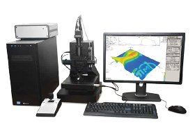
OSTEC ARTTOOL LIMITED COMPANY
Russia
IMOS Interference Microscope-nanoprofilometer enables accurate, quantitative, ISOcompliant noncontact surface measurements and characterization of micro and nanoscale surface characteristics, capturing up to two million data points in seconds. Choosing the right optical profilometer depends on your application requirements, including speed, accuracy, vertical range, automation and flexibility. IMOS optical surface profilometer provides powerful versatility in noncontact optical surface measurements. The system makes it easy and fast to investigate a wide range of surface types, including smooth, rough, flat, sloped and stepped surfaces. All measurements are nondestructive, fast and require no special sample preparation.
OSTEC ARTTOOL LIMITED COMPANY
Russia
Simultaneous / Multifunctional Analysis: - Raman - Luminescence - Laser Reflection & Transmission - Spectral and Polarization measurements The pinhole aperture rejects the residual scattered rays originating from any out-of-focus points on a sample. Spatial resolution less than 500 nm (Z), 200 nm (XY) Spectral resolution ~0.25 cm-1 Wavelength accuracy in spectrum with CCD detector 0.005 nm (1800 l/mm) Specially designed imaging spectrometer incorporates many features that make it ideal for confocal Raman measurements. The image of the pinhole is projected to a multichannel detector without any aberrations. People with little or no experience in Raman spectroscopy can use RAMOS N500. The system is highly modular and fully automated. Up to 5 lasers can be used. The lasers can be switched from one to another with just one click. Motorized control for laser power, beam diameter, polarization orientation, pinhole size, and grating is provided.
OSTEC ARTTOOL LIMITED COMPANY
Russia
RAMOS E/M series Raman spectrometers are designed on the basis of research-grade optical microscopes allowing the realization of the following light microscopy methods: - Raman measurements - Transmitted light - Reflected light (bright field and dark field illumination) - Confocal microscopy - Fluorescence measurements - Polarization contrast and phase-contrast imaging - Differential interference contrast 3D scanning laser Raman microscopes provide rapid, high sensitivity analysis. The innovative approach to system design of Raman spectrometers ensures extremely high temperature and temporal stability of spectral measurements. All RAMOS E200 system components are fully integrated within an optical microscope providing compactness and mobility of the system. In RAMOS M350, M520, M750 systems external imaging spectrographs are connected via optical fibers. Raman measurements with the RAMOS E/M Series systems can be started in several minutes by turning a system key.
OSTEC ARTTOOL LIMITED COMPANY
Russia
RAMOS U120 compact single-channel confocal Raman microscope is designed for micro spectral measurements with capabilities at the level of high-end systems. RAMOS U120 microscope has a rigid, moving parts free design that requires no adjustments, has both high sensitivity and high spatial resolution. A wide range of capabilities, high reliability, and compact size allow using RAMOS U120 for various scientific and industrial applications. Main features: - Research level optical microscope with advanced measurement techniques - Submicron resolution due to confocal design - Automatic adjustment of laser power - Wide dynamic range and extremely high sensitivity of innovative sCMOS detector - Edge or Notch filters for Stokes and AntiStokes spectroscopy - Automatic switching between Raman, optical and combined Raman-optical modes - Fiber optic Raman probe option - Raman mapping with motorized sample stage - Laser Safety Class 3B
OSTEC ARTTOOL LIMITED COMPANY
Russia
RAMOS S120 compact dual-channel confocal Raman microscope is designed for micro spectral measurements with capabilities at the level of high-end systems. RAMOS S120 microscope has a rigid, moving parts free design that requires no adjustments, has both high sensitivity and high spatial resolution, and can be equipped with two single-mode lasers simultaneously, 488/633 nm or 532/785 nm. Wide possibilities, high reliability, and compactness allow using RAMOS S120 for solving a wide range of scientific and industrial applications. Main features: - Research level optical microscope with advanced measurement techniques - Submicron resolution due to confocal design - One or two integrated single-mode lasers - Fully automated change of lasers/gratings without additional system alignment - Automatic adjustment of laser power - Wide dynamic range and extremely high sensitivity of innovative sCMOS detector - Fiber optic Raman probe option - Raman mapping with motorized sample stage
NT-MDT LLC
Russia
Early warning and forecasting of the deterioration of engineering materials operating in extreme conditions is a high priority activity within many industrial plants. The ability to perform diagnostics on a plant’s metal work without taking it offline is particularly of high value to industries in the energy, processing and other sectors. Currently diagnostic equipment is only able to discover deterioration in operational pipework and associated equipment after its condition has already reached a dangerous stage of degradation. SOLVER Pipe is a new highprecision and reliable diagnostic system that allows plant managers the ability to decrease risks and achieve incidentfree operation through anticipatory control and scheduled maintenance of materials and equipment within their industrial facilities.Advantage over electron microscopy compact, cheap and does not require complex vacuum technology.

NT-MDT LLC
Russia
Operating a scanning probe microscope, where the tipsample force interactions are measured with high precision and at small scales, requires an environment that is free of the external perturbations caused by vibrational and acoustic noise. Just as important is the temperature stability of the microscope in order to ensure a low thermal drift of a sample during experiments. A unique feature of Atomic Force Microscopy (AFM) and Scanning Tunneling Microscopy (STM) is a relatively slow feedback mechanism in most of their modes. Therefore, a large number of experiments such as imaging at the atomicscale, profiling of corrugated surfaces, collecting of local force curves in the force volume operation, among others will benefit from lowthermal drift conditions. A stable position of the sample will also help to examine the same surface region with different AFM modes, making the surface analysis more comprehensive.
NT-MDT LLC
Russia
Industry leading automation level Outstanding noise floor and thermal drifts Fast scanner with XYZ lownoise closeloop Routine atomic resolution 60+ SPM modes in basic configuration Continuous zoom from millimeter to nanometer range Integrated with new Atomic Force Microscopy technique HybriD Mode™ Atomic Force Microscope NEXT provides motorized sample positioning and integrated high resolution optical microscope positioning, motorized continuous zoom and focusing of the optical microscope. But AFM automation is more than just motorization. Powerful Nova PX software algorithms remove a gap between optics and AFM providing continuous zoom from huge panoramic optical view down to atomic resolution. Since all step movers are coupled together with the optical image, NEXT provides autofocus, fast oneclick cantilever alignment, panoramic optical view and multiple scanning on 5×5 mm range.Cantilever recognition and automatic laser alignment both in liquid and air Autofocus
NT-MDT LLC
Russia
IR s‑SNOM microscopy and spectroscopy with 10 nm spatial resolution Wide spectral range of operation 312 μm Incredibly low thermal drift and high signal stability Versatile AFM with advanced modes SRI (conductivity), KPFM (surface potential), SCM (capacitance), MFM (magnetic properties), PFM (piezoelectric forces) HybriD Mode™ quantitative nanomechanical mapping Integration with microRaman (optional) The ability of s‑SNOM measurements in the visible spectral range (optional) NTMDT Spectrum Instruments presents NTEGRA Nano IR scattering scanning nearfield optical microscope (s‑SNOM) designed for infrared (IR) spectral range. AFM probe is located in the focus of optical system which excites sample structure by IR laser and collects the optical response. Collected light is directed to Michelson interferometer for optical analysis.
Results for
Microscopes - Import exportNumber of results
16 ProductsCountries
Company type